
VSculpt / Men / Gynecomastia
Understanding gynecomastia begins with understanding you.
A private, practical conversation about chest fullness, what may be causing it and whether treatment is appropriate.
Book a Consultation
Does this sound like you?
The questions you may not have said aloud.
Feeling self-conscious about your chest can affect ordinary moments. There is room to discuss it without judgement.

Clothing
You find yourself thinking about how a shirt sits across your chest.
Activity
Swimming, exercise or changing in shared spaces feels uncomfortable.
Uncertainty
You have tried to understand whether the fullness is fat, glandular tissue or something else.
Understanding it
What exactly is gynecomastia?
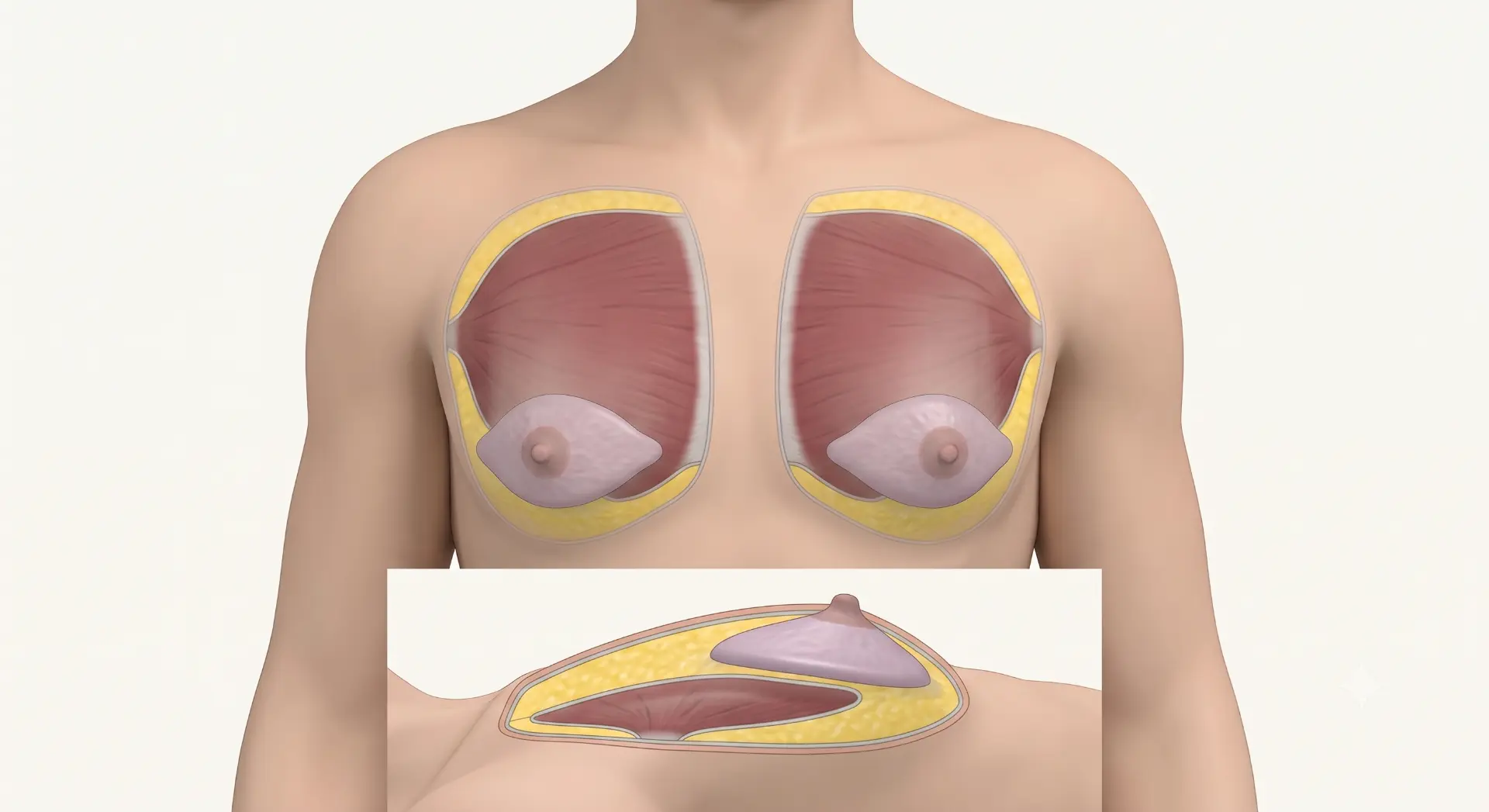
Gynecomastia describes enlargement of glandular tissue in the male chest. Chest fullness may also involve fat; a clinical assessment helps distinguish the contributors and identify whether further evaluation is needed.
- Skin — outer envelope
- Subcutaneous fat
- Gland beneath the areola
- Pectoralis muscle — behind tissue
- Ribs / chest wall — deepest
Simplified illustration, not to scale. Fat and gland lie above muscle. Gynecomastia is benign, but not every chest lump is gynecomastia.
Watch · What is gynecomastia?
What is gynecomastia?
Start here / explainer
Do I have gynecomastia?
Chest fullness is not one diagnosis. Examination can help distinguish the possible causes.
Video · Do I have gynecomastia?
A hard or irregular lump, bloody nipple discharge, nipple retraction, persistent skin changes, rapid unexplained enlargement or enlarged lymph nodes need medical assessment.
Explore the assessment →Your assessment
Begin with the right questions.
A private set of prompts to help you prepare for a conversation, not an automated diagnosis.
Private self-reflection · Question 1 of 10
How old are you?
Educational only. Not a diagnosis or a treatment recommendation.
Fat or gland?
Understanding what is causing your chest enlargement.
Move between the tissue patterns to see how a chest can differ from mostly fat to mostly gland.
Anatomy explorer
Equal fat and gland
Fatty fullness tends to be softer and more diffuse, while glandular tissue can feel firmer beneath the nipple. Many chests contain both, which is why fat distribution, gland size, skin elasticity, symmetry and nipple position are all assessed.
A visual grades guide
The same diagnosis, different chest shapes.
Grades describe enlargement and skin change. They are not instructions for an operation.
Grade I
Mild / localised
Localised fullness with little or no excess skin.
Grade IIA
Moderate
More noticeable enlargement without significant skin excess.
Grade IIB
Moderate / skin change
Fullness with some skin excess and a more apparent lower fold.
Grade III
Marked skin excess
More extensive enlargement with a looser skin envelope and lower nipple position.
Illustrative, not diagnostic. Examination determines the tissue, skin and nipple anatomy; a photograph cannot establish your grade or plan.
Watch · Grades of gynecomastia, explained
Grades of gynecomastia, explained
Watch the educational grades animation
Educational animation · the grades
Meet your surgeon
Dr. Charan J. C.
Plastic, Cosmetic & Gender-Affirming Surgeon. MBBS · MS (General Surgery) · MCh (Plastic & Reconstructive Surgery).

A useful assessment looks beyond a single label: gland, fat, skin, nipple position, symmetry and the contour you hope to achieve.
Meet Dr. Charan →Before & after
Patient photographs, shared with consent.
These images are individual cases. They illustrate what surgery can look like; they do not predict your own result.
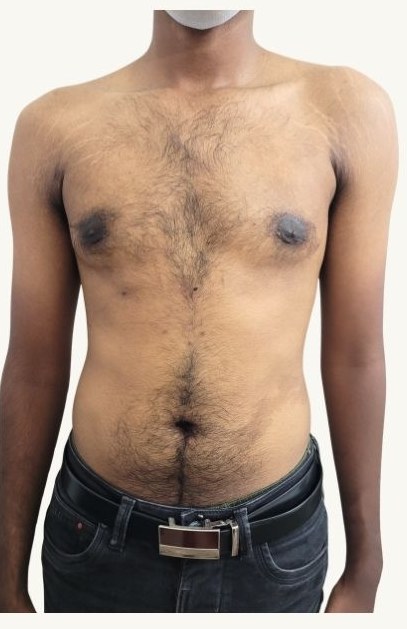
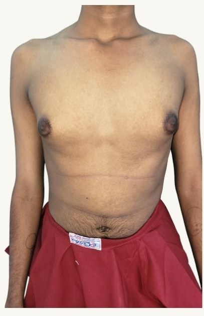
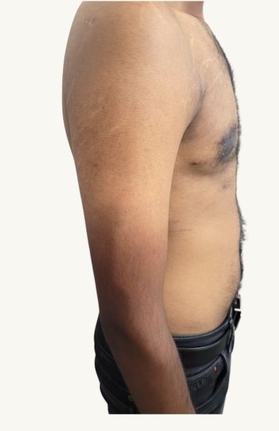
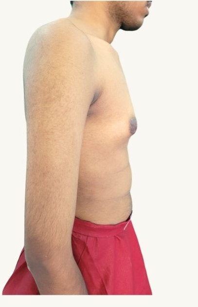
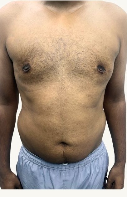
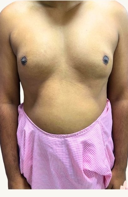
Actual patient photographs shared with consent. Outcomes differ with anatomy, grade, technique and healing.
The most common question
“I've been training my chest. Why isn't it going away?”
Exercise changes fat and muscle, but it does not behave the same way with glandular tissue.
Video · Can exercise cure gynecomastia?
Fat can reduce
Fatty fullness often decreases as overall body fat comes down with diet and training.
Gland usually stays
Glandular tissue does not behave like fat and generally does not disappear with exercise.
Muscle changes shape, not tissue
Building the chest muscle can alter the shape beneath, but it does not remove gland or overlying skin.
Why it happens
Why does gynecomastia happen?
Several factors may contribute, and extensive hormonal testing is not required for everyone.
Video · Causes of gynecomastia
Hormonal changes
Changes in hormone balance may contribute to glandular enlargement.
Puberty
Chest changes around puberty can sometimes settle without surgery; persistent or concerning changes deserve assessment.
Medication and substances
Some medicines, hormones and anabolic steroids may contribute. Review these with a clinician rather than stopping prescribed treatment yourself.
Weight and other conditions
Fat can add to chest fullness, while some medical conditions need separate evaluation.
In many men no single cause is identified, and that is not unusual. Do not stop prescribed medicine without medical advice.
Anatomy before technique
The grade doesn’t choose the surgery. Your anatomy does.
Dr. Charan assesses these factors together, then discusses a plan for your chest rather than an operation assigned to a grade.
No single factor decides treatment. Examination, your health and your priorities complete the picture.
Surgeon-led contour planning
A male chest is a contour, not simply a gland to remove.
The centre of the chest needs to relate to the surrounding chest, not be treated in isolation.
- Central gland
- Peripheral fat
- Lower chest fullness
- Lateral / axillary transition
- Skin envelope
- Nipple position
- Left–right asymmetry
- Pectoral border
The aim is a natural transition across the chest, balancing residual fullness against over-resection and a crater beneath the nipple. Perfect symmetry or a particular contour cannot be guaranteed.
Planning, safety and follow-up
Examination and measurements guide the incision and tissue removal. Your desired contour is discussed before a technique is chosen; anaesthetic fitness and medical history come first. Risks, alternatives and limits should be clear in your own words. Follow-up reviews healing, scars and contour settling.
Candidacy
Is surgery right for me?
Sometimes a medical review or more time is more useful than an operation.
You may be ready to consider surgery
Persistent fullness, stable circumstances, good general health and realistic expectations can form part of a suitability conversation.
Sometimes waiting is better
Recent puberty, unexplained changes, medication-related concerns, unstable weight or an unclear diagnosis may call for assessment first.
Video · Who can have surgery?
Video · Who should wait?
What actually changes?
How gynecomastia surgery reshapes tissue.
These are possible tools, not five steps every patient needs. Fat, gland and skin may require different approaches.
01 / Before
Before
Skin lies over subcutaneous fat and glandular tissue; pectoralis muscle and chest wall lie deeper.
02 / Liposuction
Liposuction
A cannula can reduce and feather fatty fullness. Dense gland is different tissue and may need a separate approach.
03 / Gland excision
Gland excision
Access near the areolar margin allows the dense glandular component to be addressed. The amount removed is planned carefully.
04 / Combined contouring
Combined contouring
Fat and gland are addressed together where appropriate, aiming for a gradual chest transition rather than a hollow beneath the nipple.
05 / Skin reduction
Skin reduction
Only when clinically relevant: excess skin may require additional incisions. Skin management and scar patterns vary.
Simplified and not to scale. Skin is superficial; fat and gland sit above pectoralis muscle and the chest wall. The actual plan and scars vary.
Watch · How is surgery performed?
How is surgery performed?
Incisions & scars
Will I have a scar?
Every operation leaves a scar; where it sits and how it matures is part of the plan.
Periareolar access
Liposuction access
Skin reduction, if relevant
Illustrative incision / scar concepts, not promised results. Access points and scar pattern depend on technique and anatomy.
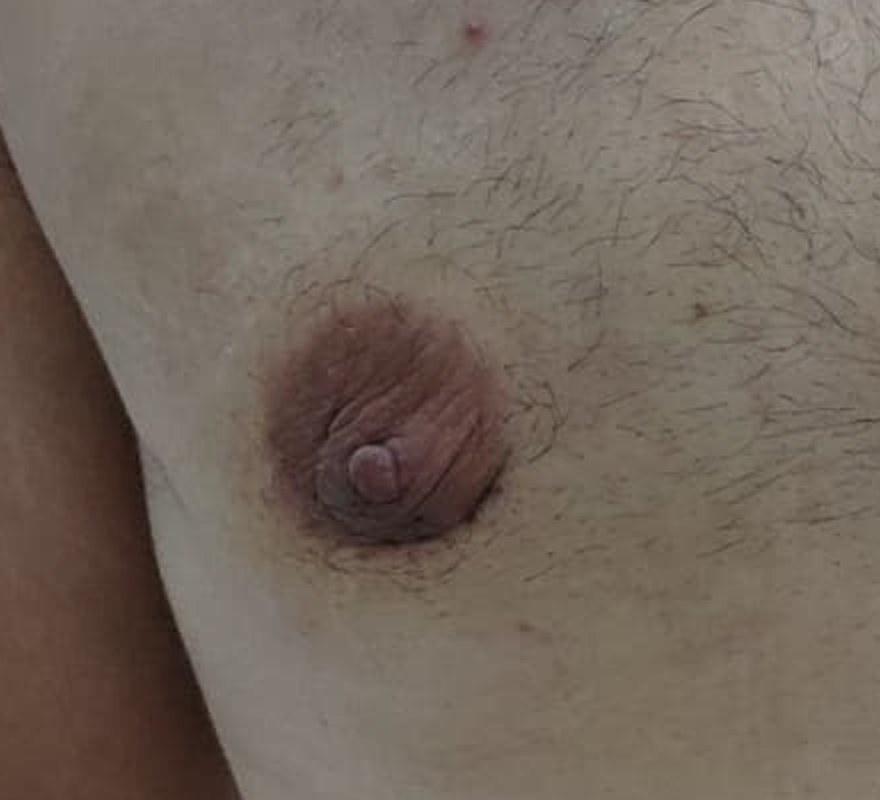
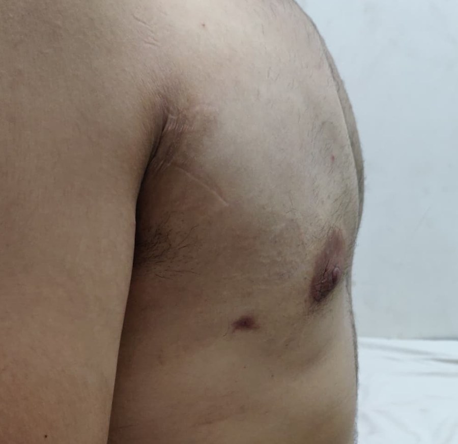
Actual patient photographs shared with consent. Your own incision plan is discussed at consultation.
All surgery leaves a scar
The aim is to place incisions as discreetly as the technique allows.
Pattern follows technique
The incision depends on the approach used and the extent of the enlargement.
Scars mature slowly
Scars typically soften and fade over many months.
Skin reduction adds scars
More extensive skin reduction requires additional incisions, discussed beforehand.
Video · Will there be a scar?
One recovery pathway
Healing moves forward in stages, not fixed dates.
Your team adapts dressings, support, reviews and activity to the operation and your healing.
- 01
Surgery & support
Dressings and chest support are explained before discharge, alongside medication, washing and sleeping advice.
- 02
Early review
Swelling, bruising and firmness can obscure the early contour. Reviews assess healing and any concerns.
- 03
Drains / sutures
If used, their care and review are arranged according to the technique and your healing.
- 04
Compression & daily life
Compression supports healing tissue; its use and duration are individual. Work, travel and driving are discussed around comfort, medication and follow-up.
- 05
Gradual activity
Light activity comes first. Lifting and exercise resume only when cleared; driving also needs safe movement and no impairing medication.
- 06
Chest exercise
Chest training is generally later in the progression, after your surgeon clears it.
- 07
Contour & scar settling
Swelling resolves and tissues soften over months. Scars and contour continue to evolve; timing and final appearance vary.
Everyday care and gradual settling
Your team explains medicines, wound care, washing, sleeping position and travel before you go home. Do not change prescribed medicines without clinician advice. Compression is used for an individually advised period and stopped after review. Work and driving depend on safe movement, the demands of your role and medication; exercise clearance is individual.
Early swelling may temporarily hide the contour. Around one month some early swelling may be settling; around three months definition may be clearer, and around six months tissues may continue softening. These are approximate, variable observations, not clearance dates or guarantees. Contour and scars can continue to change over months.
Watch · Postoperative protocol
Postoperative protocol
Watch · Restrictions and recovery
Restrictions and recovery
Watch · When will results be visible?
When will results be visible?
Common concerns
The questions men usually ask next.
Recurrence, pain and the fear of something more serious deserve direct answers.
Will gynecomastia come back after surgery?
Recurrence cannot be ruled out. Future hormonal changes, certain medicines, anabolic steroid exposure or significant weight changes may affect the chest over time.
How painful is surgery?
Most men describe soreness, tightness and tenderness rather than severe pain, particularly in the first few days. Pain relief is provided and discomfort usually eases as healing progresses.
Can gynecomastia turn into cancer?
Gynecomastia itself is a benign condition. However, not every male breast lump should be assumed to be gynecomastia, which is why examination matters.
Video · Will it come back?
Video · Will it be painful?
Video · Can it turn to cancer?
Honest information
Risks, at a glance.
Every surgical procedure carries risk. Each of these is discussed openly in consultation.
Early healing
Bleeding, infection, seroma, delayed healing and visible scarring are possible.
Contour and symmetry
Contour irregularity, asymmetry, residual fullness, over-resection and the need for revision surgery can occur.
General risks
Temporary or lasting nipple sensation change and anaesthesia-related risks are discussed in consultation.
Seek assessment promptly
A hard or irregular lump, bloody nipple discharge, nipple retraction, skin changes, rapid unexplained enlargement or enlarged lymph nodes should be examined.
Cost · safety · FAQs
What else should you know?
Questions about cost and risk deserve individual answers, not campaign promises.
Understanding the cost
The fee depends on assessment, technique, anaesthesia, facility and individual care needs. A personalised estimate can be discussed after consultation; no price is promised here.
The next step is yours
A clearer understanding starts with a conversation.
Bring your questions. We can discuss what is happening, your options and whether treatment is right for you.